

Your retina is the light-sensitive tissue at the back of your eye. Retinal detachment is a medical emergency that occurs when this tissue is pulled away from its normal position. If retinal detachment goes untreated for a long period of time, your risk of permanent vision loss in that affected eye is greater. Early treatment of retinal detachment is crucial to protecting your vision
Retinal detachment is painless but does come with warning signs and symptoms before it occurs. These can include:
- Shadows over your field of vision
- Blurred vision
- The appearance of floaters
- Photopsia (flashes of light in either one or both of your eyes)
- Reduced peripheral vision
Retinal Detachment Surgery
The type of surgery you require depends on where in your eye it detached and much of your retina is detached. There are three types of surgery that can be performed to fix a detached retina. You will be advised on the type of surgery recommended for your particular case. In some instances, you may need more than one surgery at once. The types of surgery include:
- Vitrectomy – small instruments are used to cut the vitreous (the gel-like fluid that fills your eye) and suction it out.
- Scleral Buckle – your eyeball will be exposed and a freezing instrument may be used to seal your retina. A small device called a scleral buckle can be used to hold your retina in place.
- If it is affecting your vision, it can be treated with vitrectomy surgery.
- Pneumatic retinopexy – an expanding gas bubble will be injected into your eye. This bubble floats over the detached area and pushes it against the back of your eye. Your retina will then be sealed against the wall of your eye.
Epiretinal Membrane Surgery
An epiretinal membrane is a thin sheet of fibrous tissue found on the inner surface of the retina. Also known as macular fibrosis or a macular pucker, it develops as a result of cellular changes that occur in the back of your eye. If your epiretinal membrane is mild and has little or no effect on your vision, then surgery will not be necessary. If it is affecting your vision, Dr Vayanos will treat it with vitrectomy surgery.
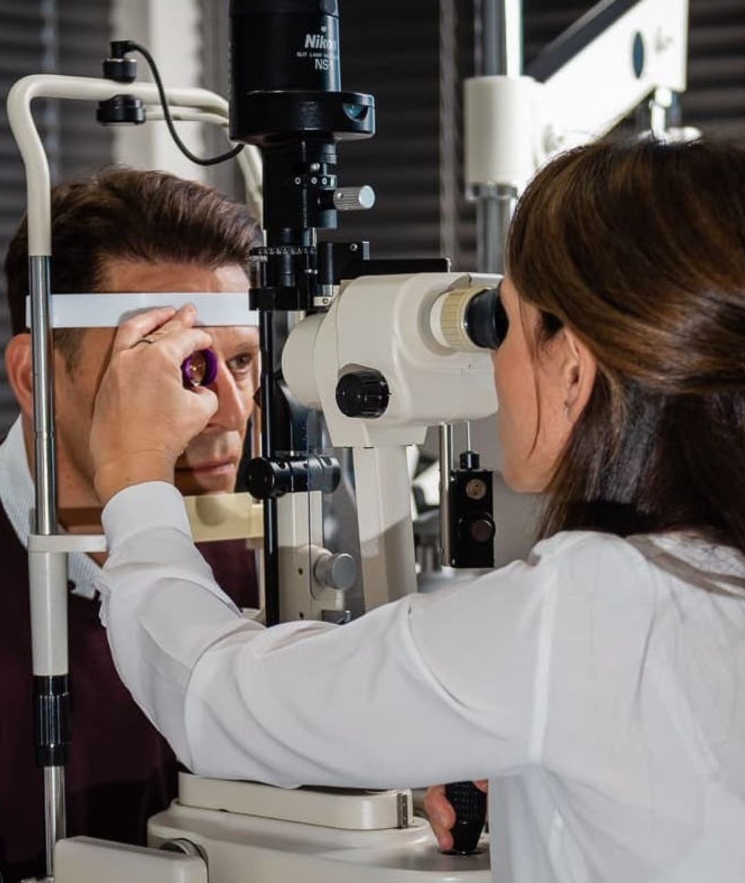
Macular Hole Surgery
A macular hole is a small gap or break at the centre of your retina. This area is called the macula. In some cases, a macular hole will seal itself and require no treatment or surgery. A macular hole happens when the retina tears. This occurs when the vitreous is firmly attached to the retina when it pulls away.
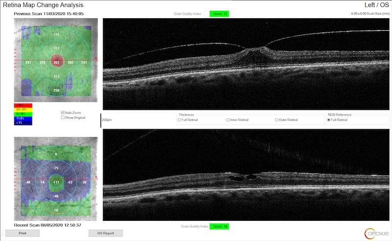
Macula holes occur more often in females and patients over 60. It is not entirely understood why they these holes occur. Dr. Vayanos will use a technology called Optical Coherence Tomography or OCT to confirm the presence of a macular hole. This non-invasive imaging technology is used to obtain cross-sectional images of your retina. Often, Macular Holes can be repaired using vitrectomy surgery.
Floaterectomy
Floater vitrectomy or floaterectomy for short, is a procedure used to remove the vitreous (gel-like fluid) from your eye. Eye floaters are those annoying spots or specks that drift about when you move your eyes. These spots in your vision are typically due to the aging of the vitreous gel.
For many patients, these spots can be nothing but a minor annoyance but for some, they can be potentially dangerous. When floaters pop up into your field of vision while driving for instance, they can obscure your vision leading to an accident.
If floaters have become a major nuisance, Dr Vayanos will evaluate you and consider floaterectomy. She will discuss any issues and concerns you may have and help you to make an informed decision regarding surgery.
During surgery, the vitreous will be removed through a small incision and will be replaced with a solution to help your eye maintain its shape. After surgery, your eye may be red, swollen or tender. This usually lasts several weeks and full recovery after a floaterectomy is usually between 2 and 4 weeks.
There are always potential risks with any eye surgery and the same goes for floaterectomy. Fortunately, severe complications are very rare but they can include retinal detachment, haemorrhage, and infection. If floaters are greatly affecting the quality of your life, Dr. Vayanos recommends you consider surgery as soon as possible.
Ophthalmologist in Cape Town
Dr Vayanos is a general Ophthalmologist in full-time private practice at the Netcare Christiaan Barnard Private hospital in the centre of Cape Town, South Africa.
Her practice is equipped with the latest technology allowing her to give you personalised, quality, and high-standard eye care and surgical precision. One of her areas of expertise includes surgical retina services.
Have any questions on retinal surgery? We are here to help. If you do not find your answer below, get in touch with us and we will do our best to provide it.
Before returning to your normal activities, you will need 2-4 weeks to recover from retinal detachment surgery.
Retinal surgery generally boasts a high success rate, with vision preservation being achievable for most patients. However, some individuals may experience a recurrence of retinal detachment, necessitating two or more surgical interventions. Common risks associated with all retinal surgeries include bleeding and infection.
You typically will not experience pain during retinal surgery. However, during the recovery period, some discomfort may be felt, and the eye may appear swollen and red.
Possible complications of retinal surgery depend on the type of procedure you receive. These can include infection, cataracts and glaucoma.
You will remain awake throughout the procedure. Bright lights will be noticeable, and some minor discomfort may be experienced. A local anaesthetic will be administered via injection into the tissues surrounding the eye to numb the area.
Initially, coping with the consequences of retinal detachment can be challenging. Treatment options are available, and their effectiveness often hinges on the timeliness of intervention and the extent of existing damage. However, following retinal detachment surgery, most individuals are able to maintain a good level of vision and live independently.
As one ages, the vitreous humour in the eye may undergo changes in texture and shrinkage. In some cases, this shrinkage can cause the vitreous to pull on the retina, potentially leading to a tear. Other factors that can increase the risk of rhegmatogenous retinal detachment include eye injuries, prior eye surgeries, and nearsightedness.
While the surgery is often very successful, complications can occur. The possible complications and side effects of retinal detachment surgery include intraocular pressure, infection, bleeding, cataract formation and epiretinal membrane.