

A lot of the human brain is involved in vision and visual processing. Neuro-ophthalmic conditions are vision problems that relate to your nervous system. These neurological disorders can have varied ocular symptoms that range from painful eye movement to dimming of vision and even permanent visual loss.
Dr Vayanos is an experienced ophthalmologist that deals with neurologic disorders that affect your vision. Disturbances in your vision can be caused by various disorders specific to the eye. These include:
Optic neuritis
Optic neuritis is a common neuro-ophthalmic condition. It occurs when inflammation or swelling damages your optic nerve. These nerves transmit visual information from your eye to your brain. Multiple sclerosis is a disease that is most commonly associated with optic neuritis.
Causes of optic neuritis
Although the exact cause of optic neuritis is not always clear, it is believed to be a fault in the immune system. It targets and attacks the protective cover of your optic nerve (myelin) resulting in a damaged and inflamed myelin. Other causes of optical neuritis include bacterial and viral infections such as Lyme disease and mumps and measles. Autoimmune diseases such as lupus and medications such as antibiotics are also believed to cause the condition.
Symptoms of optic neuritis
- Dimming of vision – usually in one eye
- Pain – eye pain that is worsened by eye movement
- Flashing lights – or flickering lights when you move your eye
- Colour vision loss – colours will appear less vivid than normal
- Visual field loss – peripheral and central vision loss can occur
Consult Dr Vayanos as soon as possible should you have any unusual symptoms, if you develop new symptoms, or if your symptoms should worsen. She will conduct a few tests that could include all or some of the following:
- A routine eye exam – this is done to check your colour vision, your peripheral vision, and a visual acuity test (to determine the smallest letters you can read on a chart)
- An MRI (magnetic resonance imaging) – this test creates a detailed image of your brain allowing Dr Vayanos to look for any damaged areas
- Pupillary reaction test – a bright light will be shone in front of your eyes. This is done to check how they respond
- Ophthalmoscopy – this is done to check if your optic nerve is swollen
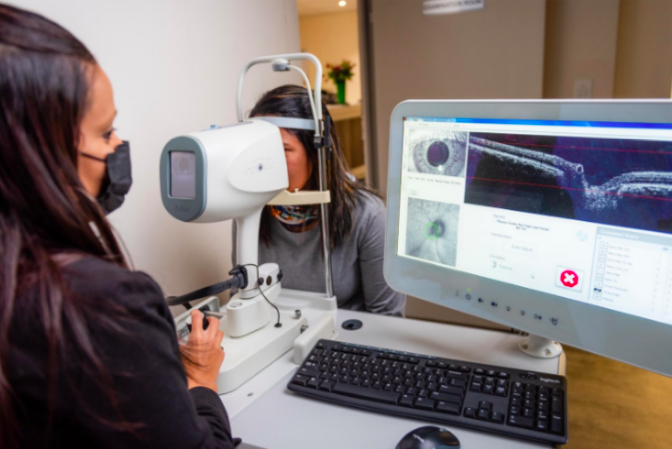
- OCT (optical coherence tomography) – this is done to measure the fibre layer in your retinal nerve
- VER (visual evoked response) – electrodes will be applied to your head to measure the speed at which your optic nerve sends signals to your brain
Treatment for optic neuritis
- High-dose steroid drugs through an IV
- IVIG (intravenous immune globulin)
- Vitamin B12 injections
Ischemic Optic Neuropathy
Ischemic optic neuropathy (ION) occurs when blood does not flow properly to your optic nerve. It can cause lasting damage to the nerve and result in sudden loss of vision in one or both of your eyes. Ischemic optic neuropathy can affect you central or peripheral and in some cases, both. Optic nerve damage cannot be fixed which means permanent vision loss can occur from ION.
Symptoms, Diagnosis & Treatment
Before ION begins, you will experience a TIA (transient ischemic attack) Your vision will darken for a few seconds or minutes and then return to normal. This is caused by reduced blood flow to your optic nerve. If you experience a TIA, consult with Dr Vayanos immediately. High-risk patients include those with high blood pressure, diabetes, and glaucoma. If you are a smoker, have clogged arteries, or are over the age of 50, you are also at risk of developing ION.
Dr Vayanos will conduct an eye exam to look out for signs of ION. She will dilate your pupils with eye drops to check for optic nerve swelling. She may also measure the fluid pressure in your eye and test your peripheral vision. If you are diagnosed with ION, treatment can include steroid pills or medication to control your diabetes, high blood pressure, or any of the other health issues mentioned above.
Glaucomatous Optic Neuropathy
Glaucomatous optic neuropathy (GON) is a feature of glaucoma in the optic nerve that causes loss of peripheral vision and in severe untreated cases, eventual blindness. GON is characterised by progressive loss of RGCs or retinal ganglion cells. All types of glaucoma involve glaucomatous optic neuropathy and these can be diagnosed and treated by Dr Vayanos.
Neuro Ophthalmologist in Cape Town
Dr Vayanos is a general Ophthalmologist in full-time private practice at the Netcare Christiaan Barnard Memorial Hospital in the centre of Cape Town, South Africa. One of her areas of expertise includes neuro-ophthalmological problems.
Her practice is equipped with the latest technology allowing her to give you personalised, quality, and a high-standard of eye care and surgical precision. If you have experienced any of the symptoms mentioned above, we recommend you pay a visit to Dr Vayanos as soon as possible.

Have any questions on neuro-ophthalmic conditions? We are here to help. If you do not find your answer below, get in touch with us and we will do our best to provide it
Neuro-ophthalmologic disease encompasses a range of visual disorders related to the optic nerve, eye movements, and the brain regions responsible for vision.
Optic neuritis often resolves on its own within 4 to 12 weeks, with vision improving as the inflammation diminishes. In severe or persistent cases, high-dose steroid drugs through an IV may be administered to speed up recovery.
Most patients with optic neuritis typically experience eye pain that intensifies with eye movement, often described as a dull ache behind the eye. Vision loss in one eye is common, with most people experiencing some degree of temporary vision reduction, although the severity of this loss can vary.
Optic neuropathy results from damage to the optic nerve and can arise due to various factors. These include infections such as toxoplasmosis and herpes simplex, inflammatory diseases, neurological disorders, certain medications, dietary deficiencies, and exposure to toxins like methanol, alcohol, and tobacco.
Optic neuritis refers to the inflammation of the optic nerve, which transmits visual information from the eye to the brain. While optic neuritis is often linked to multiple sclerosis (MS), it does not necessarily indicate that a person has or will develop MS. Many individuals experience optic neuritis without any subsequent symptoms.
Signs of optic nerve damage include a relatively sudden decrease in vision, which may manifest as blurring, darkening, or dimming. Vision loss affecting the centre, a portion, or the entirety of the visual field. Abnormalities in colour perception, such as dull or faded colours. Pain in or around the eye, often exacerbated by eye movement.
Typically, the fluid within the eye (aqueous humor) circulates freely through the anterior chamber and exits via the drainage system known as the trabecular meshwork. However, if this drainage system becomes blocked or malfunctions, intraocular pressure increases, leading to optic nerve damage.
Current treatment approaches for Primary Open-Angle Glaucoma (POAG) and Normal-Tension Glaucoma (NTG) focus on reducing intraocular pressure (IOP) through the use of topical ocular medications, combination drug therapies, and surgical procedures.